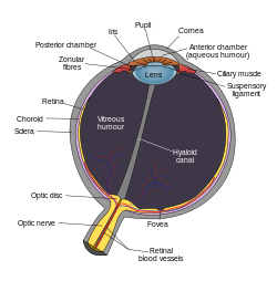
朱令案眼科问题的焦点是外眼下毒是否能够直接扩散到达视网膜。其等价问题就是滴眼液进入内眼之后如何分布。让我们首先来了解一下必要的解剖和生理知识。Nile引用的知识来自PharmaTutor网站,文章标题是《 REVIEW ON OCULAR DRUG DELIVERY 》。这是网上可以找到的最详尽的专业资讯。有关滴眼液在内眼的分布和走向全文如下: After eye drop administration the peak concentration in the anterior chamber is reached after 20–30 min, but this concentration is typically two orders of magnitude lower than the instilled concentration even for lipophilic compounds [21]. From the aqueous humor the drug has an easy access to the iris and ciliary body, where the drug may bind to melanin. Melanin bound drug may form a reservoir that is released gradually to the surrounding cells, thereby prolonging the drug activity. Distribution to the lens is much slower than the distribution to the uvea [22]. Unlike porous uvea, the lens is tightly packed protein rich structure where drug partitioning takes place slowly. Drug is eliminated from the aqueous humor by two main mechanisms: by aqueous turnover through the chamber angle and Sclemm’s canal and by the venous blood flow of the anterior uvea [22] (Fig. 3). The first mechanism has a rate of about 3μl/min and this convective flow is independent of the drug. Elimination by the uveal blood flow, on the other hand, depends on the drug’s ability to penetrate across the endothelial walls of the vessels. For this reason, clearance from the anterior chamber is faster for lipophilic than for hydrophilic drugs. Clearance of lipophilic drugs can be in the range of 20–30 μl/min. In those cases, most of drug elimination takes place via uveal blood flow. Halflifes of drugs in the anterior chamber are typically short, about an hour. The volumes of distribution are difficult to determine due to the slow equilibration of drug in the ocular tissues. The estimates in rabbits range from the volume of aqueous humor (250 μl) up to 2 ml [23]. In the latter case, the slow drug distribution to the vitreous is included in the volume of distribution. This distribution is slow, because the lens prohibits drug access to the vitreous. Flow of aqueous humor from the posterior chamber to the anterior chamber is another limiting factor.
http://www.pharmatutor.org/articles/review-on-ocular-drug-delivery?page=0,2 这段文章的关键知识点在这里:
• peak concentration in the anterior chamber is reached after 20–30 min
• drug has an easy access to the iris and ciliary body
• Distribution to the lens is much slower than the distribution to the uvea, the lens is tightly packed protein rich structure where drug partitioning takes place slowly.
• most of drug elimination takes place via uveal blood flow
• vitreous distribution is slow, because the lens prohibits drug access to the vitreous 
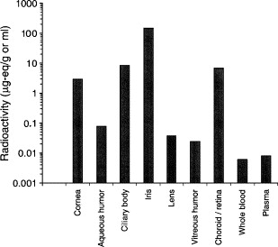
滴眼液进入前房后随房水从后房向前房的循环回流进入脉络膜静脉。这是经得起反复检验也很容易理解的科学事实。药物在内眼的分布晶状体是一个重要的限制因素,也可以看作是药物在内眼的分水岭。在晶状体前面的,虹膜(iris)和睫状体(ciliary body)药物有比较高的分布。到了晶状体,情况就变了:the lens is tightly packed protein rich structure where drug partitioning takes place slowly. vitreous distribution is slow, because the lens prohibits drug access to the vitreous。很清楚,也很容易理解,药物不仅很难进入晶状体而且还会被晶状体阻挡难以进入其身后的玻璃体。 现在特别注意这句:Distribution to the lens is much slower than the distribution to the uvea。什么是uvea。Uvea=The vascular middle layer of the eye constituting the iris, ciliary body, and choroid. iris, ciliary body前面都提到了,那么这里的choroid又是什么。Choroid=The dark-brown vascular coat of the eye between the sclera and the retina. Also called choroid coat, choroid membrane. 具备了这些知识,我们来一起解读一张图。这张图来自是万维著名骗子pzzdm的托儿travel1提供的一篇用放射性物质研究药物在内眼分布的论文。自从nile把他们的骗局当场揭穿,这位骗子就不遗余力查文献用来反驳nile所说的滴眼液不能直接扩散到达视网膜。现在我们就来认真研究一下他提供的这份数据。  Fig. 4. Radioactivity in ocular and systemic tissues of cynomolgus monkeys after 5 days of topical 0.2% [14C]brimonidine tartrate b.i.d. Concentrations of radioactivity were measured at 2 h after the last dose of drug. Mean values are shown (n = 4 eyes). Fig. 4. Radioactivity in ocular and systemic tissues of cynomolgus monkeys after 5 days of topical 0.2% [14C]brimonidine tartrate b.i.d. Concentrations of radioactivity were measured at 2 h after the last dose of drug. Mean values are shown (n = 4 eyes).
这张条图清楚表明,药物透过角膜进入内眼后,浓度分布从低到高是iris, ciliary body 和aqueous humor。再往下依次是晶状体和玻璃体。其中的浓度比iris低上千倍,与全血浓度比高不过十倍。这与我们上面了解到的知识是吻合的。由于房水循环清除药物和晶状体对药物扩散的屏障,从虹膜到玻璃体药物浓度已经下降了1000倍。现在问题来了。在choroid/retina 中的浓度与ciliary基本一样,高于玻璃体数百倍。这是为什么? 其实原因我们已经知道了。“Distribution to the lens is much slower than the distribution to the uvea.”。choroid(葡萄膜)是uvea(脉络膜)的一个重要组成部分,相当于相机中的暗箱用来屏蔽光线。前面已经提到,uvea包括iris, ciliary body, and choroid。药物在uvea上分布比晶状体多得多。这就是为什么choroid/retina 的药物浓度与ciliary基本一样的原因。choroid/retina中的药物浓度是choroid中药物浓度贡献的,与retina基本没有关系。视网膜前面的玻璃体药物浓度已经降到了内眼最低,视网膜没有从内眼来源的药物可以使其药物浓度突然上升数百倍。如果能把retina单独分离下来测定,其中的药物浓度应该低于晶状体和玻璃体,与全血浓度持平。也就是说非常接近平均全血浓度。 骗子travel1对眼球的解剖生理一无所知。不了解系统知识当然会对他煞费苦心查到的论文只知其一不知其二。所以误打误撞,以为他查到的论文给nile的打击是“毁灭性”的。不料想事与愿违,这篇论文为早已成熟的眼球解剖生理和药物动力学知识提供了一个有力的佐证。骗子们不断卖弄你们的无知和愚蠢即在情理之中,也不出意料之外。但是科学以实证为根据建立的理论不会因为你们的需要作出改变。你们急于洗清骗子臭名的心情nile可以理解。唯一的方法就是公开谢罪,不是向nile而是向所有的网友!
|
